Welcome!
I am the Chief Medical Physics Resident in the Department of Radiation Oncology at Indiana University, where I focus on the clinical aspects of cancer treatment through radiation. My research interests include radiation physics, radiopharmaceutical therapy, and Monte Carlo simulations. I specialize in applying Monte Carlo simulations to address challenges in radiation physics, encompassing dosimetry and radiation biology. A key feature of my research is close collaborations with experts in radiopharmaceutical dosimetry, molecular imaging, and radiation transport simulations.
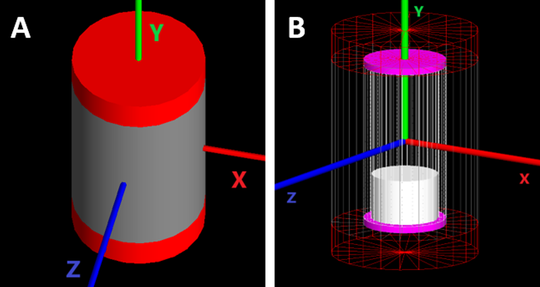
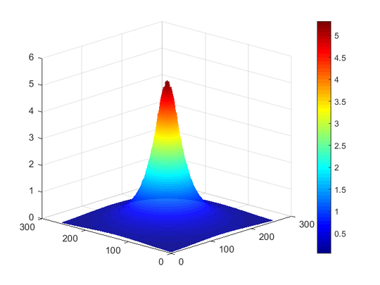
My doctoral thesis explored dosimetry in radiopharmaceutical therapy and quantitative imaging in nuclear medicine. Specifically, I investigated into two main areas: (1) dosimetry of β- and α-emitting radionuclides, and (2) Monte Carlo simulation of PET scanners, complemented by rigorous validation.
Short Bio:
I hail from Nareshwor, Gorkha, Nepal, where I cherished my formative years. I earned my B.Sc. (2008) and M.Sc. (2012) degrees from Tribhuvan University. I then pursued my second master’s degree at the University of South Dakota in 2017 and completed my Ph.D. in physics at the University of Iowa in 2022.
Outside of my professional life, I find joy in cycling and appreciate the tranquility of nature. My literary interests include both fiction and non-fiction, and I also enjoy various gaming activities, such as Carrom, card games, and exploring culinary arts.
- Radiation Physics
- Radiopharmaceutical Therapy
- Monte Carlo Simulations
- Positron Emission Tomography
- Theranostics
-
Certificate in Medical Physics, 2023
Wake Forest University, Winston-Salem, NC, USA
-
PhD in Physics, 2022
University of Iowa, Iowa City, IA, USA
-
MS in Physics, 2017
University of South Dakota, Vermillion, SD, USA
-
MSc in Physics, 2012
Tribhuvan University, Kathmandu, Nepal
-
BSc in Physics, 2008
Tribhuvan University, Kathmandu, Nepal
Skills
90%
100%
90%
100%
100%
70%
Experience
Responsibilities include:
- QA for patient-specific treatment plans and machines (daily/monthly/annual)
- Performed QA for linacs, Gamma Knife, and HDR systems
- Treatment planning: VMAT/IMRT, 3D (RapidArc/HyperArc)
- Teaching Therapy Physics to residents
- Involved in clinical physics research
Responsibilities include:
- Research on radiation biology using Ac-225 radioisotope
- Investigation on PET imaging extravasation dosimetry
Responsibilities include:
- Research
- Mentor undergraduate student
Responsibilities include:
- Graduate Research Assistant (Fall 2018 - Spring 2022): Conducted research and authored a PhD thesis
- Graduate Teaching Assistant (Fall 2017 - Spring 2018): Assisted in physics labs and graded lab reports
Responsibilities included:
- Graduate Research Assistant (Spring 2016 - Spring 2017)
- Graduate Teaching Assistant (Fall 2015)
- Assisted studens in lab, graded their lab reports
- Authored MS thesis
Responsibilities include:
- Taught introductory nuclear phyiscs to class XII students
- Taught heat and thermodynamics to class XI students
- Designed exams, assited in physics labs, and graded assignments
Projects
Recent & Upcoming Talks
Featured Publications
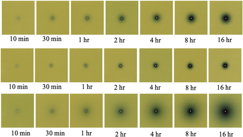
The aim of this project is to experimentally validate the Monte Carlo generated absorbed doses from the beta particles emitted by 90Y and 177Lu using radiochromic EBT3 film‐based dosimetry.
Line sources of 90Y and 177Lu were inserted longitudinally through blocks of low‐density polyethylene and tissue‐equivalent slabs of cortical bone and lung equivalent plastics. Radiochromic film (Gafchromic EBT3) was laser cut to accommodate orthogonal line sources of radioactivity, and the film was sandwiched intimately between the rectangular blocks to achieve charged particle equilibrium. Line sources consisted of plastic capillary tube of length (13 ± 0.1) cm, with 0.42‐mm inner diameter and a wall thickness of 0.21 mm. 90Y line sources were prepared from a solution of dissolved 90Y resin microspheres. 177Lu line sources were prepared from an aliquot of 177Lu‐DOTATATE. Film exposures were conducted for durations ranging from 10 min to 38 h. Radiochromic film calibration was performed by irradiation with 6‐MV‐bremsstrahlung x rays from a calibrated linear accelerator, in accordance with literature recommendations. Experimental geometries were precisely simulated within the GATE Monte Carlo toolkit, which has previously been used for the generation of dose point kernels.
The mean percentage difference between measured and simulated absorbed doses were 5.04% and 7.21% for 90Y and 177Lu beta absorbed dose in the range of (0.1–10) Gy. Additionally, 1D gamma analysis using a local 10%/1 mm gamma criterion was performed to compare the absorbed dose distributions. The percentage of measurement points passing the gamma criterion, averaged over all tests, was 93.5%. We report the experimental validation of Monte Carlo derived beta absorbed dose distributions for 90Y and 177Lu, solidifying the validity of using Monte Carlo‐based methods for estimating absorbed dose from beta emitters.
Overall, excellent agreement was observed between the experimental beta absorbed doses in the linear region of the radiochromic film and the GATE Monte Carlo simulations demonstrating that radiochromic film dosimetry has sufficient sensitivity and spatial resolution to be used as a tool for measuring beta decay absorbed dose distributions.
Recent Publications
Contact
- tiwarias@yahoo.com
- 605-202-1567
- 838 Blake Street Apt I, Indianapolis, IN 46202
- DM Me
