Monte Carlo evaluation of hypothetical long axial field-of-view PET scanner using GE Discovery MI PET front-end architecture
Abstract
Purpose: The development of total-body PET scanners is of growing interest in the PET community. Investigation into the imaging properties of a hypothetical extended axial field-of-view (AFOV) GE Healthcare SiPM-based Discovery MI (DMI) system architecture has not yet been performed. In this work, we assessed its potential as a whole-body scanner using Monte Carlo simulations. The aim of this work was to (1) develop and validate a Monte Carlo model of a four-ring scanner and (2) extend its AFOV up to 2 m to evaluate performance gain through NEMA-based evaluation.
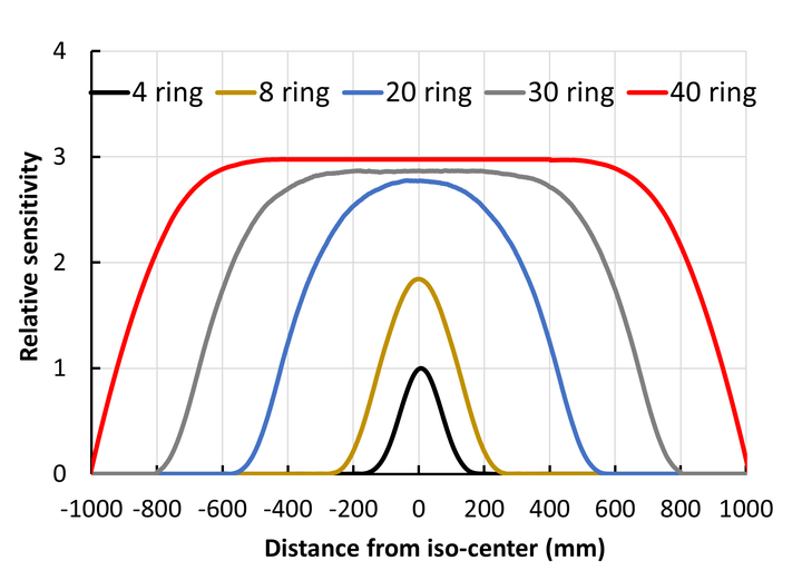
Methods: The DMI four-ring geometry and its pulse digitization scheme were modeled within the GATE Monte Carlo platform using published literature. The GATE scanner model was validated by comparing results against published NEMA performance measurements. Following the validation of the four-ring model, the model was extended to simulate 8-, 20-, 30-, and 40-ring systems. Spatial resolution, sensitivity, NECR, and scatter fraction were characterized with modified NEMA NU-2 2018 standards; however, the image quality measurements were not acquired due to computational limitations. Spatial resolutions were simulated for all scanner ring configurations using point sources to examine the effects of parallax errors. NEMA count rates were estimated using a standard 70 cm scatter phantom and an extended version of scatter phantom of length 200 cm with (1-800) MBq of 18F for all scanners. Sensitivity was evaluated using NEMA methods with a 70 cm standard and a 200 cm long line source.
Results: The average FWHM of the radial/tangential/axial spatial resolution reconstructed with filtered back-projection at 1 and 10 cm from the scanner center were 3.94/4.10/4.41 mm and 5.29/4.89/5.90 mm for the four-ring scanner. Sensitivity was determined to be 14.86 cps/kBq at the center of the FOV for the four-ring scanner using a 70 cm line source. Sensitivity enhancement up to 21-fold and 60-fold were observed for 1 and 2 m AFOV scanners compared to four-ring scanner using a 200 cm long line source. Spatial resolution simulations in a 2 m AFOV scanner suggest a maximum degradation of ∼23.8% in the axial resolution compared to the four-ring scanner. However, the transverse resolution was found to be relatively constant when increasing the axial acceptance angle up to ±70°. The peak NECR was 212.92 kcps at 22.70 kBq/ml with a scatter fraction of 38.9% for a four-ring scanner with a 70 cm scatter phantom. Comparison of peak NECR using the 200 cm long scatter phantom relative to the four-ring scanner resulted in a NECR gain of 15 for the 20-ring and 28 for the 40-ring geometry. Spatial resolution, sensitivity, and scatter fraction showed an agreement within ∼7% compared with published measured values.
Conclusions: The four-ring DMI scanner simulation was successfully validated against published NEMA measurements. Sensitivity and NECR performance of extended 1 and 2 m AFOV scanners based upon the DMI architecture were subsequently simulated. Increases in sensitivity and count-rate performance are consistent with prior simulation studies utilizing extensions of the Siemens mCT architecture and published NEMA measurements with the uEXPLORER system.