Large Chest Phantom for Positron Emission Tomography
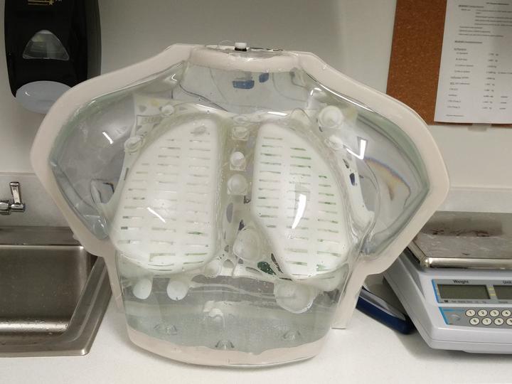 Final product of the phantom
The aim of this work is to make the large chest anthropomorphic phantom that closely mimicks the average american adult (male) chest.
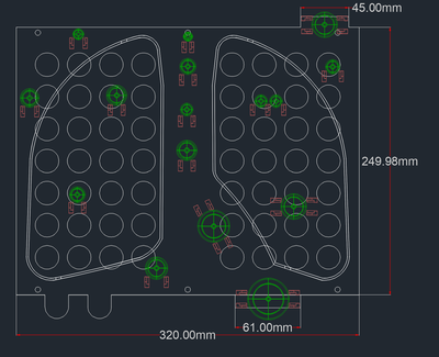
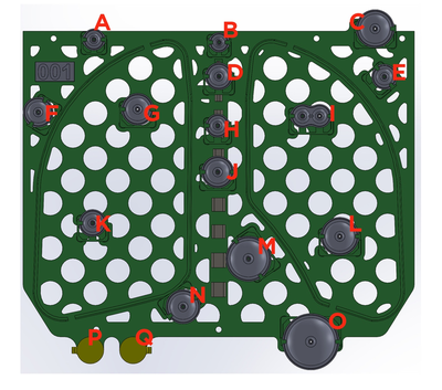
Interior of the Phantom:
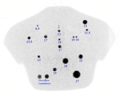
There are a total of 16 spheres (tumors) inside the phantom. This includes 12 Clinical Trial Network (CTN) chest phantom spheres (37, 28, 22, 22, 17, 17, 13, 13, 10, 10, 10, 7 mm) and four additional spheres of internal diameters (19, 15.5, 11.5, and 8.5 mm). These dimensions are the interior diameter of the spheres. All spheres and interior platform of the phantom were 3D printed. Each sphere has a shell thickness of 1 mm. The 12 CTN size speres are placed in the same position as that in CTN chest phantom for fair comparison. The lung cavities consists of spongy materials that closely mimicks the lung tissue of density ~0.3 $ \frac{g}{cm^{3} }$. The total volume occupied by the lungs in this phantom is ~6.038 Liter.
Phantom measurements:
- Dry phantom weight = 3.54 kg
- Phantom fill volume = 22.35 Liter
- Total phantom weight after filling = 25.90 kg
Ashok Tiwari, PhD
Medical Physics Resident
My research interests include Radiation Physics, Nuclear Medicine, Monte Carlo Simulations and Artificial Intelligence.